Orthodontic Treatment for Sleep Apnea: Clinical Mechanisms and Structural Airway Solutions
Obstructive sleep apnea (OSA) is a sleep-related breathing disorder characterized by repeated episodes of partial or complete upper airway collapse during sleep. These nocturnal obstructions lead to recurrent oxygen desaturations, fragmented sleep architecture, and progressive strain on the cardiovascular system. While Continuous Positive Airway Pressure (CPAP) therapy remains the traditional first-line medical intervention, long-term patient compliance frequently falters due to interface discomfort, mask leaks, and claustrophobia.
At Dentovex, our clinical team approaches upper airway obstruction from a structural and dentofacial perspective. Modern airway-centric orthodontics does not view the dental arches in isolation; it views the hard tissues of the jaws as the structural scaffolding for the upper respiratory tract. By modifying the transverse, sagittal, and vertical relationships of the maxilla and mandible, orthodontic treatment for sleep apnea addresses root anatomical contributors to pharyngeal collapse.
Craniofacial Phenotypes and Airway Resistance
Pharyngeal collapsibility during sleep is heavily governed by the surrounding bony architecture. Soft tissue structures, including the soft palate, uvula, lateral pharyngeal walls, and tongue base, are suspended directly or indirectly from the skull base, maxilla, and mandible. When skeletal discrepancies reduce the internal dimensions of this bony container, the soft tissues are displaced posteriorly into the pharyngeal airway space.
Clinicians identify several skeletal phenotypes that predispose patients to sleep-disordered breathing:
- Mandibular Retrognathia: A retruded lower jaw repositions the genioglossus muscle and tongue base closer to the posterior pharyngeal wall, significantly narrowing the retroglossal airway space.
- Transverse Maxillary Deficiency: A narrow, high-arched hard palate encroaches directly upon the nasal vault. This elevates nasal airway resistance, encouraging chronic mouth breathing. Mouth breathing allows the mandible to drop down and back, further precipitating upper airway collapse.
- Bimaxillary Retrusion: Hypoplasia of both jaws reduces total oral cavity volume, forcing normal-sized soft tissue structures into a restricted space.
Re-establishing stable skeletal dimensions through orthodontic and dentofacial orthopedic techniques can permanently enlarge these critical anatomical spaces.
Primary Orthodontic Modalities for Airway Management
Depending on patient age, skeletal maturity, and the severity of the Apnea-Hypopnea Index (AHI), orthodontists utilize several targeted modalities to alleviate airway restriction.
1. Custom Mandibular Advancement Splints (MAS)
For adult patients with mild to moderate obstructive sleep apnea, custom oral appliance therapy serves as the primary non-surgical orthodontic intervention. Unlike non-custom, over-the-counter boil-and-bite devices, a clinical mandibular advancement splint is fabricated from precision digital impressions or physical polyvinyl siloxane models.
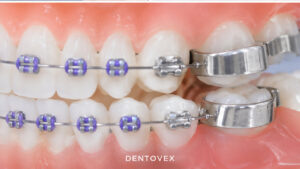
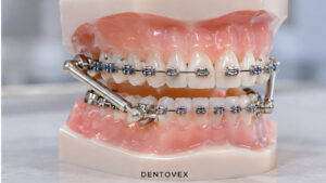
These split-arch appliances engage the upper and lower teeth to advance the mandible into a protrusive, forward position during sleep. This mechanical translation pulls the tongue base forward via the lingual attachments, tenses the soft palate through the palatoglossus muscle, and stabilizes the lateral pharyngeal walls against negative inspiratory pressures. Appliances are incrementally titrated until optimal airway patency is reached without inducing excessive strain on the temporomandibular joints (TMJ).
2. Maxillary Skeletal Expansion (MSE / MARPE)
In patients exhibiting transverse maxillary constriction and elevated nasal resistance, expanding the midpalatal suture yields profound changes in upper airway dimensions. In growing pediatric and adolescent patients, traditional Rapid Palatal Expansion (RPE) reliably opens the suture. In skeletally mature adults, orthodontists utilize Miniscrew-Assisted Rapid Palatal Expansion (MARPE).
By securing an expansion framework directly to the palatal vault with micro-implants, MARPE applies orthopedic forces directly to the basal bone, bypassing dental tipping. This separates the midpalatal suture, widens the floor of the nasal cavity, increases internal nasal volume, and reduces nasal resistance, which facilitates natural closed-mouth nasal breathing during sleep.
3. Surgical Orthodontics: Maxillomandibular Advancement (MMA)
For severe OSA cases characterized by profound skeletal deformities or where non-invasive therapies have failed, combined orthodontic-surgical treatment offers definitive correction. Maxillomandibular Advancement (MMA) involves Le Fort I osteotomy of the maxilla paired with bilateral sagittal split osteotomy (BSSO) of the mandible. The jaws are advanced anteriorly, typically by 10 to 12 millimeters [VERIFY]. This structural movement tensions the entire muscular framework of the pharynx, expanding the velopharyngeal, retroglossal, and hypopharyngeal airways simultaneously.
Comparative Overview of Airway-Targeted Interventions
The following table contrasts the biomechanical parameters, patient demands, and primary clinical applications of the predominant orthodontic and dental interventions against conventional therapy.
| Clinical Parameter | Mandibular Advancement Device (MAD) | Miniscrew-Assisted Expansion (MARPE) | Maxillomandibular Advancement (MMA) | CPAP Therapy (Medical Standard) |
|---|---|---|---|---|
| Primary Target Site | Retroglossal and retrovelar pharynx | Nasal floor and anterior palatal vault | Global pharyngeal airway (naso-, oro-, and hypopharynx) | Entire upper respiratory tract (pneumatic) |
| Mechanism of Action | Mechanical mandibular protrusion via dentition | True skeletal transverse expansion via bone-anchored screws | Skeletal advancement of maxilla and mandible via osteotomies | Positive pneumatic pressure splinting soft tissues |
| Invasiveness | Non-invasive, reversible nocturnal wear | Minimally invasive (local anesthesia for micro-implants) | Major inpatient surgical procedure under general anesthesia | Non-invasive, requires continuous nocturnal mask wear |
| Typical Target Group | Mild-to-moderate OSA; CPAP-intolerant patients | Skeletally mature adults with narrow maxilla and nasal stenosis | Severe refractory OSA; pronounced skeletal retrognathia | Moderate-to-severe OSA across all anatomical presentations |
| Reported Compliance | High to moderate (65% to 80% long-term wear) [VERIFY] | Fixed intraoral appliance; compliance is passive | High (permanent structural alteration; no nocturnal wear required) | Moderate to poor (40% to 50% non-adherence rate) [VERIFY] |
The Clinical Pathway for Orthodontic Airway Management
Treating sleep-disordered breathing within an orthodontic framework requires a multidisciplinary workflow alongside board-certified sleep medicine physicians.
- 1. Medical Diagnostics and Polysomnography
The patient completes an overnight diagnostic sleep study (polysomnography or an approved home sleep test) interpreted by a board-certified sleep physician to establish baseline AHI, oxygen desaturation indices, and a formal diagnosis of OSA.
- 2. Orthodontic and Cephalometric Assessment
The orthodontic team evaluates the craniomaxillofacial anatomy using low-dose Cone Beam Computed Tomography (CBCT). Airway cross-sectional areas, nasal patency, tongue posture, and temporomandibular joint health are systematically measured.
- 3. Appliance Selection and Precise Bite Registration
If oral appliance therapy is indicated, a George Gauge or digital bite registration captures the target therapeutic position, typically starting at 60% to 70% of the patient maximum comfortable mandibular protrusion [VERIFY].
- 4. Appliance Insertion and Controlled Titration
The custom device is delivered, verified for uniform retention and occlusal stability, and advanced incrementally over several weeks to balance airway improvement against muscular and joint adaptation.
- 5. Objective Efficacy Verification
Once clinical symptoms stabilize, a follow-up sleep study is performed with the appliance in situ to objectively verify the reduction in AHI and normalization of oxygen levels.
Critical Considerations: Temporomandibular Joint and Occlusal Health
Orthodontic management of sleep apnea is not without risks to the stomatognathic system. Because mandibular advancement splints transfer forward forces directly to the anchoring dentition and the temporomandibular joints, distinct biological adaptations can occur over extended use:
1. Occlusal Drift: Prolonged wear can lead to a slight reduction in overjet and overbite, mesial movement of lower molars, and retroclination of upper incisors. To counter these changes, patients must use a morning occlusal repositioner (a rigid morning aligner) for 10 to 15 minutes each day to help reset the mandible into centric occlusion.
2. Temporomandibular Disorders (TMD): Transient muscle soreness in the lateral pterygoid and masseter muscles is relatively common during initial titration. However, pre-existing structural internal derangements of the TMJ (such as non-reducing disc displacement) require careful clinical management or may preclude protrusive therapies entirely.
3. Periodontal and Dental Anchoring Capacity: Adequate periodontal support and a minimum number of healthy, stable teeth per arch are mandatory to distribute mechanical loads without causing tooth loosening or unwanted tipping.
Financial and Insurance Considerations
The financial investment for orthodontic management of sleep apnea varies significantly based on appliance complexity and diagnostic protocols. Custom mandibular advancement splints typically range between $1,800 and $4,200 [VERIFY], including construction, clinical insertions, and initial titration visits. Miniscrew-assisted skeletal expanders generally range from $2,500 to $5,500 [VERIFY].
Because OSA is classified as an underlying medical condition rather than a cosmetic dental issue, custom oral appliances may be covered in part under major medical insurance policies rather than dental plans, provided specific diagnostic thresholds and CPAP intolerance criteria are met. Clinicians must provide documented diagnostic sleep studies, a physician prescription, and clear treatment notes to facilitate pre-authorization.
Long-Term Prognosis and Stability
When patients are selected based on precise anatomical diagnostics, orthodontic treatment for sleep apnea offers a clinically validated, durable therapy. Skeletal expansions and surgical corrections deliver permanent changes to the airway perimeter, often significantly reducing lifelong reliance on external nocturnal appliances. For patients using custom oral advancement splints, long-term adherence rates consistently outperform CPAP therapy, translating to meaningful reductions in daytime hypersomnolence, lower systemic blood pressure profiles, and reduced cardiovascular mortality over the life course.
Medical Disclaimer:
The content provided in this article is for informational and educational purposes only and does not constitute professional medical or dental advice, diagnosis, or treatment. Always seek the advice of a qualified dentist, orthodontist, or other authorized healthcare provider regarding any medical condition or treatment plan. Never disregard professional medical advice or delay seeking it because of something you have read on the Dentovex blog.
No responses yet